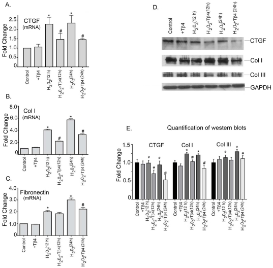
Figure 4. Effect of Tβ4 on profibrotic genes under oxidative stress in cardiac fibroblasts.
Cells were treated with H2O2 in the presence and absence of Tβ4 and (A) CTGF, (B) Col-I and (C) Fibronectin mRNA expression was analyzed at 12 h and 24 h, respectively by qRT-PCR. Data represent the means±SE of three separate experiments. (D) Western blot analysis showed the protein expression of CTGF, Col-I and Col-III at 12 h and 24 h, respectively. GAPDH was used as a loading control for the experiment. (E) Graph shows the relative fold change in the protein expression of Bax, Bcl2 and caspase-3, respectively by densitometry. Data represent means±SEM from 3 individual experiments. * denotes p<0.05, compared to controls while # denotes p<0.05 compared to the H2O2-treated group and ø denotes p>0.05, compared to controls.

