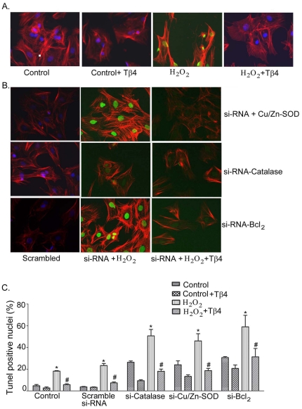
Figure 6. Effect of Tβ4 on cardiac fibroblast apoptosis under oxidative stress.
(A) Representative fluorescent microscopy images of TUNEL staining in rat neonatal cardiac fibroblasts. Bright TUNEL-positive staining (FITC) was observed in H2O2 treatment which was not observed in control cells and cells pretreated with Tβ4. DAPI was used to stain the intact nuclei and counterstaining of filamentous actin was done with Texas Red®-X phalloidin. (B) Representative fluorescent microscopy images showing the effect of Tβ4 treatment in presence and absence H2O2-induced oxidative stress on cardiac fibroblasts transfected with siRNAs of Cu/Zn-SOD, catalase and Bcl2 vs. scrambled siRNA, respectively. (C) Bar graph shows the percent TUNEL-positive nuclei under similar experimental condition. Data represent the means±SE of at least three separate experiments. A total of 65 to 82 nuclei were counted for each observation. * denotes p<0.05 compared to controls while # denotes p<0.05, compared to the H2O2-treated group.

