
Fig. 17.
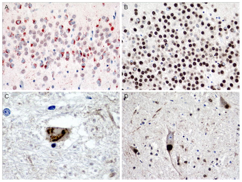
Case 3D. Comparison of NCIs in dentate gyrus (a, b) and SLIs and LBLIs in hypoglossal nucleus (c, d) in case 3C (a, c) compared to brother (b, d). Note frequent granular NCIs in case 3 with bvFTD (a) but only sparse NCIs in brother with clinical ALS (b). Additionally, hypoglossal SLIs were rare in case 3 (c), but LBLIs were frequent (and SLIs sparse, not shown) in brother (d). p-TDP-43 antibody (a), ×400 magnification, TDP-43 antibody (b, d), ×400 magnification and c ×600 magnification