
Figure 2. Directional migration is impaired in primary cilia deficient Tg737orpk/orpk endothelial cells.
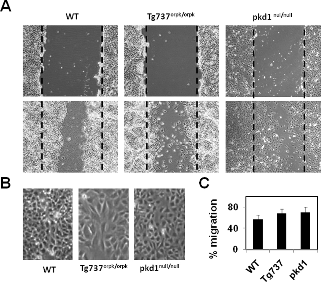
A) Photmicrographs of endothelial cells at 0 and 24 hours after wounding. The dashed line denotes the wound edge. B) Images of endothelial cells showing the orientation at the wound closure. Note: Wild type endothelial cells migrated and closed the wound by orienting perpendicular to the direction of the wound where as Tg737orpk/orpk cells oriented abnormally (parallel) to the wound. C) Quantitative analysis of the wound closure presented as % migration. The results shown are mean ± SEM from 3 independent experiments.