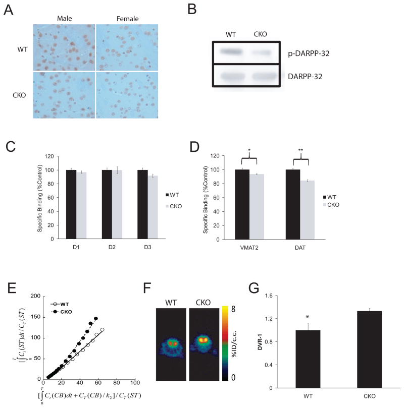
Fig. 1. Nf1 CKO mice with attention system defects demonstrate a presynaptic DA defect, which can be visualized by PET imaging.
(A) IHC reveals decreased DARPP32 phosphorylation (p-DARPP32) in the striatum of both male and female mice relative to WT littermates (p=.01; N=8). (B) Western blot shows a 5.8-fold decrease in p-DARPP32 (following normalization to total DARPP32 levels) in CKO compared to WT mice. In vitro quantitative receptor autoradiography demonstrates no change in postsynaptic D1, D2 and D3 DA receptor expression in CKO mice relative to control WT littermates (C), whereas presynaptic VMAT2 and DAT expression is reduced (D; ~10%; p=.03, VMAT2, p=.0004, DAT; N=6). (E) Representative Logan plots for WT and CKO mice (using the cerebellum as the reference region) are shown along with (F) representative [11C]-raclopride transverse micro-PET images (summed across 5–60 minutes). The colorscale bar indicates the normalized peak uptake (percent injected dose per cubic centimeter tissue; %ID/cc). (G) In a cohort of WT and CKO mice, [11C]-raclopride binding was increased in the striatum (Str) of CKO mice compared to control WT littermates on PET imaging (p=.03; N=4 per genotype). Ct = tissue radioactivity at time t; T = time point of each frame of PET scanning course.

