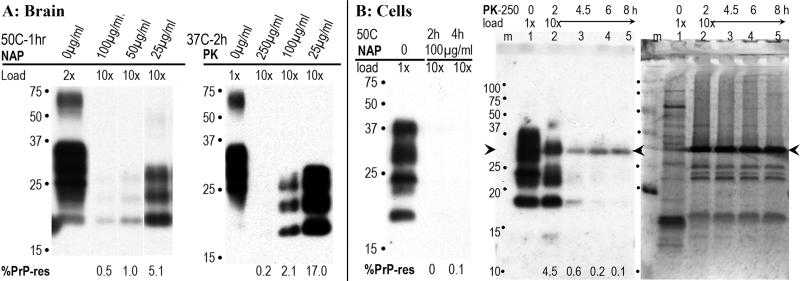
Fig. 2.
Representative digestion of brain (A) and cells (B) under different digestion conditions. In brain, different concentrations of NAP were tested for 1hr and compared with no enzyme controls (0μg/ml). At 100μg/ml 99.5% of the PrP was digested and can be seen as faint bands in the 10x gel loaded lane. A 2hr incubation with NAP did not increase digestion of PrP (data not shown). With PK, 99.8% digestion of brain PrP required 250μg/ml for 2hr. Panel B shows NAP digested almost all the PrP of FU-CJD cells at 2hr, i.e., was slightly more effective than in brain. Longer 4h digests did not further reduce the PrP (0-0.1% by photon counting) as noted. In contrast to brain, 250μg/ml PK left 4.5% PrP-res at 2hr, and longer incubations were tested as shown. It required 6-8hr to digest PrP to 0.1-0.2%. The PK enzyme in this blot bound PrP antibody (arrowhead) and corresponds to the PrP band seen with colloidal gold staining. Remarkably, exhaustive PK digestion left many obvious non-PrP protein bands between 15-29kd. The 6 and 8hr PK cell digests shown here were titered for infectivity.

