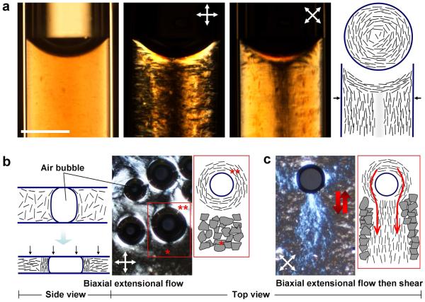
Figure 2. Assembly modes in graphene oxide aqueous phases: surface anchoring, microconfinement, and flow ordering.
a, Infiltration of 10 mg/ml GO suspension into 1 mm diameter cylindrical capillaries leads to a well-defined concentric layer patterns driven by homeotropic surface anchoring at the glass walls and shear flow into the capillary. b, Biaxial extensional flow (squeezing flow) orients GO platelets perpendicular to the compression direction leading to large non-birefringent (black) regions under crossed polars in top view (see *). The presence of air bubbles in the GO suspension also leads to vertically aligned GO layers in the near-surface region of the bubbles reflecting homeotropic surface anchoring. This alignment is clearly seen by the bright lenses at 45°/135°/225°/315° under 0°/90° crossed polars (see **). Rotation of crossed polars causes equivalent rotation in these features clearing indicating the surface anchored pattern shown in the top view sketch at the far right. c, Following biaxial extension flow by oscillating shear flow leads to vertically aligned “wakes” of GO layers forced to flow around the lens-shaped air bubbles (top view). The vertically aligned GO layers are trapped in the viscous LC phase are stable after oscillating shear flow is stopped (scale bar: 400 μm).

