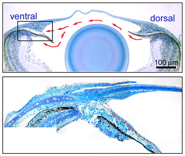
Figure 2. Iridocorneal angle of the zebrafish eye.

Histology showing low magnification of the adult zebrafish anterior segment (Upper panel). Red arrows show the general flow of aqueous humor. Higher magnification shows the openings of the ventral canalicular outflow pathway (Lower panel).
