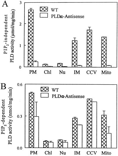
Figure 1.
Subcellular distribution of PIP2-independent (A) and -dependent (B) PLD activities in Arabidopsis leaves. The assay conditions for the two types of PLD activities were as described in Methods. Identical methods were used to isolate the intracellular fractions from fully expanded leaves of wild-type (WT) and PLDα antisense Arabidopsis. PM, Plasma membrane; Chl, chloroplast; Nu, nucleus; IM, intracellular membrane; CCV, clathrin-coated vesicle; Mito, mitochondria of wild-type and PLDα antisense plants.

