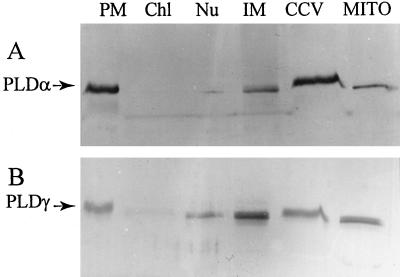
Figure 3.
Immunoblot of PLDα (A) and PLDγ (B) in various subcellular fractions. Intracellular fractions were isolated from wild-type Arabidopsis leaves, and equal amounts of proteins (10 μg per lane) were loaded and separated by 8% SDS-PAGE. PLDα and PLDγ were made visible by alkaline phosphatase after blotting with affinity-purified PLDα and PLDγ antibodies, respectively. See Figure 1 for abbreviations.

