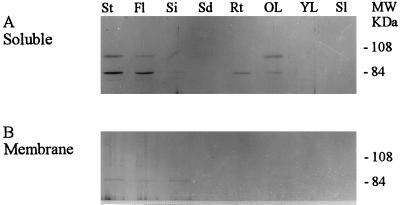
Figure 7.
Immunoblot of PLDγ in soluble (A) and membrane (B) fractions of different tissues. Equal amounts (10 μg per lane) of soluble and membrane-associated protein were separated by 8% SDS-PAGE. PLD bands were immunodetected with incubation of the filters with affinity-purified PLDγ antibody. The protein samples and abbreviations are the same as those in Figure 4.

