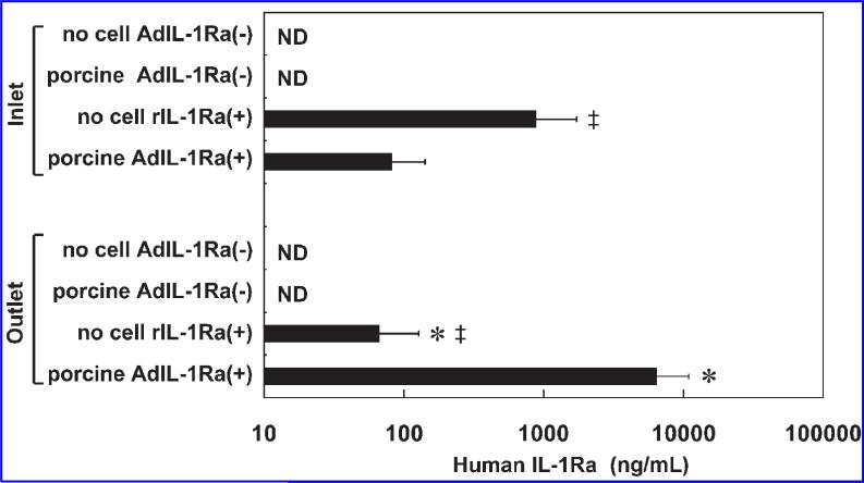
FIG. 5.
Human IL-1Ra levels at the inlet and outlet of the BAL device at the end of 10 h of extracorporeal perfusion. The results are expressed as means ± SD. *p < 0.05 vs. the same group (no cell rIL-1Ra(+) or porcine AdIL-1Ra(+)) at the inlet; ‡p < 0.05 vs. porcine AdIL-1Ra(+) group in the same group (inlet or outlet); no cell AdIL-1Ra(–), no cells in the BAL device; porcine AdIL-1Ra(–), non-transfected porcine hepatocytes in the BAL device; no cell rIL-1Ra(+), BAL device without cells, but recombinant human IL-1Ra was continuously infused through a venous line; porcine AdIL-1Ra(+), porcine hepatocytes transfected with adenoviral vector encoding human IL-1Ra. IL-1Ra, interleukin-1 receptor antagonist; BAL, bioartificial liver; ND, not detected.

