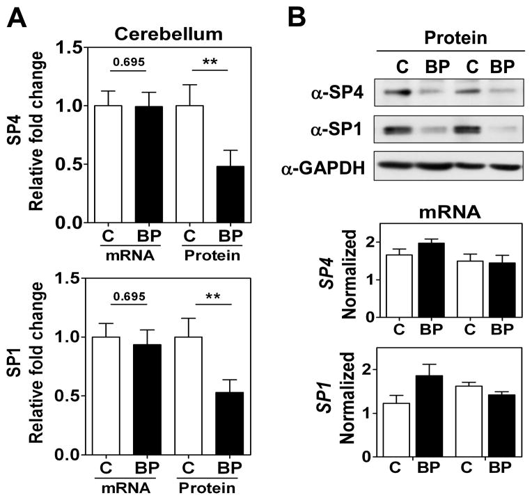
Fig. 1.
SP4 and SP1 protein levels are reduced in postmortem cerebellum in bipolar disorder (BD) subjects. (A) Protein and mRNA levels for SP4 (top) and SP1 (bottom) in the cerebellum. Protein extracts from cerebellar postmortem tissue of healthy individuals (C, n = 10), and individuals with BD (n = 10) were immunoblotted for SP4, SP1, and glyceraldehyde-3-phosphate dehydrogenase (GAPDH). The resultant bands were quantified by densitometry. SP4 and SP1 were normalized to GAPDH values and referred to a standard sample (healthy subject). Each value represents the mean of two independent analyses. mRNA levels for SP4 and SP1 from the same subject samples were determined RT-qPCR and normalized to a reference healthy control sample and the geometric mean of three reference genes: beta glucuronidase (GUSB), TATA-binding protein (TBP), and fibroblast growth factor 1 (FGF1). Each value represents the mean of at least three independent analyses performed in duplicate. Statistical analysis was performed using Wilcoxon signed rank test for paired values (**p < 0.01). (B) Representative Western blots images for SP4, SP1, and GAPDH from four individuals (top), and representative RT-qPCR data for the same four individuals (bottom).

