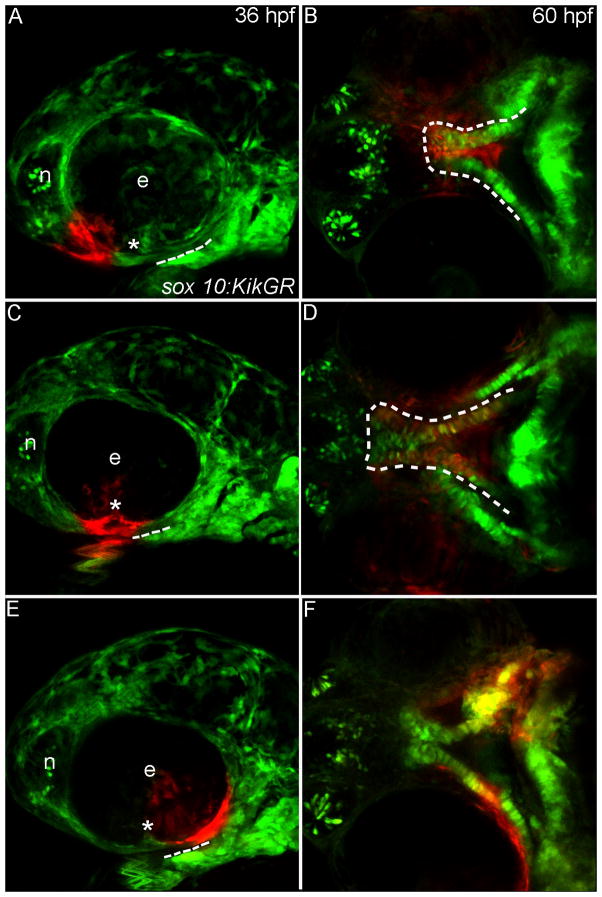
Fig. 3. Progenitor domains for the zebrafish palate.
(A–F) Sox10:KikGR photoconverted CNCC in red. (A,C,E) Lateral images of the zebrafish head at 36 hpf dashed lines indicate the oral ectoderm, asterisk marks location of the choroid fissure. (B,D,F) Ventral view of the developing palatal skeleton at 60 hpf, the developing palate is outlined by the dashed white lines. In some cases other CNCC-derived tissues were labeled as the UV penetrated through the embryo, but here we focus on the palatal skeleton. (A) CNCCs adjacent to the eye and nasal epithelium (termed frontonasal CNCC) were labeled. (B) Labeled cells occupy the midline of the ethmoid plate. (C) CNCCs adjacent to and anterior to the choroid fissure (anterior maxillary CNCC) were labeled. (D) Photoactivated CNCC populate the lateral ethmoid and anterior portion of the trabeculae. (E) CNCCs labeled posterior to the choroid fissure (posterior maxillary CNCC) (F) will contribute to the posterior portion of the trabeculae. e, eye; n, nasal opening.

