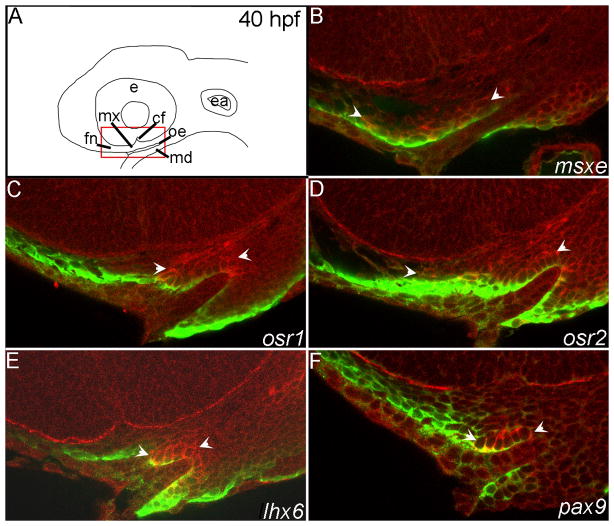
Fig. 5. Fluorescent in situ hybridization optical sections verify the expression patterns of select transcription factors at 40 hpf.
(A) Schematic diagrams of a lateral view of the zebrafish head at 40 hpf, red box indicates the magnified view of the in situ sagittal sections. (B–F) mRNA localization detected by fluorescence of the NBT/BCIP precipitate (red) and anti-EGFP immunostaining labels CNCC in the fli1:EGFP transgenic background (green). Arrowheads indicate areas of CNCC expression domains. (B) msxe is expressed in frontonasal and maxillary CNCC. (C) osr1 is expressed in maxillary CNCC. (D) osr2 is expressed in the frontonasal and maxillary CNCC. (E) Maxillary CNCC express lhx6. (F) The posterior maxillary CNCC express pax9. e,eye; mx, maxillary domain; md, mandibular domain; fn, frontonasal CNCC; cf, choroid fissure; oe, oral ectoderm; ea, ear.

