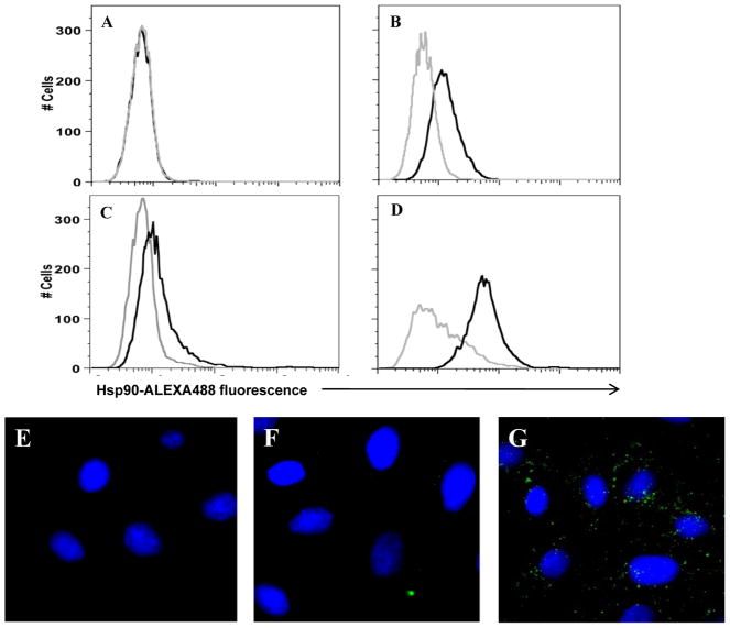
Figure 1. KSHV-permissive cells express extracellular Hsp90.
(A–D) To identify cell surface localization of Hsp90 (csHsp90) by flow cytometry, HeLa (A–C) and primary dermal microvascular endothelial cells (pDMVEC) (D) were incubated with antibodies recognizing either the C-terminus (A), N-terminus (B and D), or an uncharacterized epitope (C) of the alpha isoform of Hsp90 followed by secondary antibodies conjugated to ALEXA-488 (black histograms). For controls, cells were incubated with secondary antibodies alone (gray histograms). (E–G) To validate these results, HeLa cells incubated with secondary antibodies conjugated to ALEXA-488 (E), anti-C-terminal Hsp90 Ab and secondary antibodies (F), or anti-N-terminal Hsp90 Ab and secondary antibodies (G) were examined by fluorescence microscopy. Original magnification × 60. Data shown represent one of three independent experiments.

