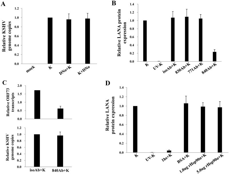
Figure 4. Direct KSHV interaction with extracellular Hsp90 is not required for KSHV entry.
(A) HeLa cells were incubated with media alone (mock), KSHV alone (K), 1.0 μM DNo 16 h before KSHV (DNo+K) or for 16 h beginning immediately following KSHV incubation (K+DNo), and qPCR was used to determine relative intracellular KSHV DNA content in the above groups. (B) Cells were incubated with KSHV alone, UV-KSHV alone, or 30 μg/mL monoclonal antibodies recognizing either the N-terminus (771Ab) C-terminus (830Ab), or an uncharactierized epitope (840Ab) of Hsp90 or isotype control Ab (isoAb) for 12 h prior to incubation with KSHV. LANA expression was quantified by IFA 12 h after viral incubation. (C) Cell-free KSHV pellets were incubated with 1.0 mg/mL of heparan sulfate (hs) for 1.5 h at 37°C, 1.0 μg BSA, 1.0 μg or 5.0 μg purified recombinant Hsp90-α for 1 h at 37°C, or media alone prior to infection. LANA expression was quantified by IFA 12 h later as above. Error bars represent the S.E.M. for three independent experiments.

