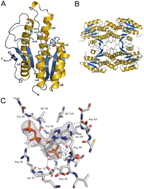
Figure 5. The crystal structure of BpiB09 in complex with NADP+.
A) BpiB09 monomer, β1–7 indicate the β sheets and α 1–6 indicates the alpha helices. Catalytic residues are indicated together with the NADP-binding site residues. B) Tetrameric arrangement of BpiB09 as probably present in solution together with the bound cofactor NADP. C) Cofactor binding in BpiB09. Supposed hydrogen bonds are shown as orange broken lines. The electron density at the NADP molecule is contoured at 1 σ.

