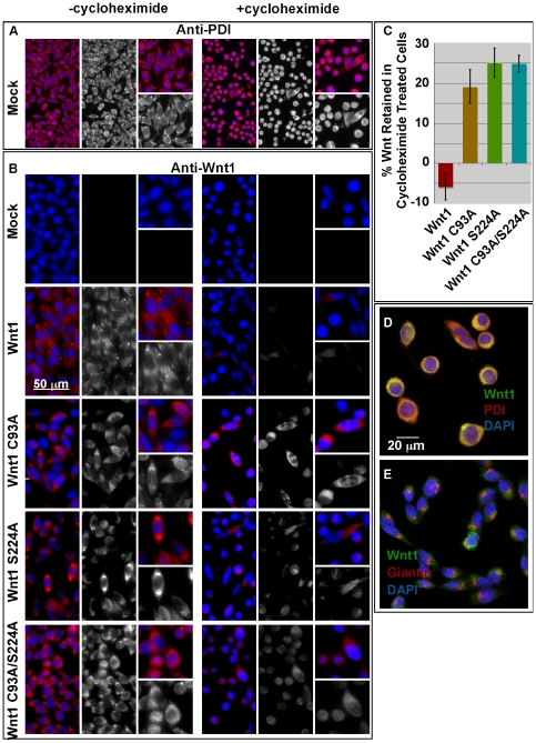
Figure 4. Mutation of the cysteine and/or serine causes an accumulation of Wnt protein in the endoplasmic reticulum.
Stably transfected L cells were grown in the presence (+cycloheximide) and absence (-cycloheximide) for 14–21 hours. Cells were then fixed and stained for PDI (Panel A) or Wnt1 (Panel B). Images were collected with a SPOT camera attached to a Nikon E600 compound microscope. A scale bar is shown for reference. This experiment was carried out twice. Panel C: For quantitation of these studies, images were collected via confocal microscopy. Fields for imaging were selected while visualizing only the DAPI channel. NIH ImageJ was used to measure the cell intensities in the Wnt1 channel. The data shown represents the sum of 3 independent replicates with a total of 9 fields counted (3 for each replicate). At least 120 cells were counted for each data point. Error bars indicate +/− standard error. In Panels D and E, confocal analysis of Wnt1 C93A/S224A of cycloheximide treated cells is shown. Cells were immunostained with anti-Wnt1 (green), anti-PDI (red; Panel D), and anti-Giantin (red; Panel E). DAPI (blue) was used to visualize the nuclei.

