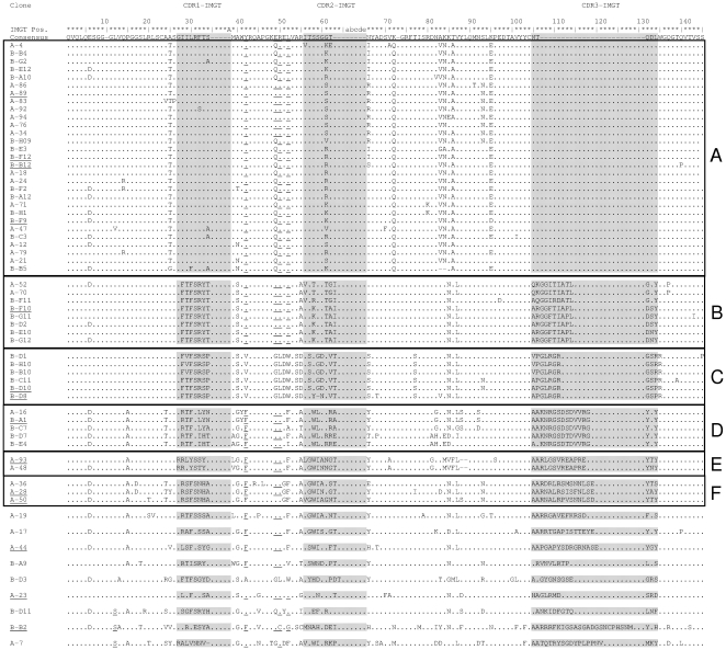
Figure 2. M. tuberculosis binding VHH antibody fragments.
Protein sequence of 62 selected VHH antibody fragments selected by phage display for binging to M. tuberculosis. Dots indicate sequence identity, and dashes indicate gaps. The three complementarity determining regions CDR1, CDR2 and CDR3 are shaded. Characteristic VH-VHH hallmark substitutions (Leu12Ser, Val42Phe or Val42Tyr, Gly49Glu, Leu50Arg or Leu50Cys and Trp52Gly or Trp52Leu (the last substitution is less well conserved) (the ImMunoGeneTics system [52] was followed) [15] are underlined. The 12 clones selected for further investigations are underlined. VHH protein sequences labeled A-x (with x = 1–96) resulted from direct selection using semi-purified protein antigen, protein sequences labeled B-yx (with y = A–F and x = 1–12) were achieved by depletion method.

