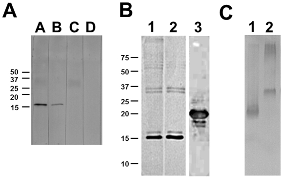
Figure 4. Western blot to discover antigen of VHHs.
A) Western blot using VHH A-23 as a probe. 9 µg M. tb 1 lysate were run on a 15% SDS-PAGE gel in lanes A–D, and transferred to a nitrocellulose membrane. Lane A: incubated with VHH A-23 and detected using anti-VSV-HRP; Lane B: incubated with VHH A-23 and detected using anti-HIS-HRP; Lane C: incubated with detection antibody anti-VSV-HRP; Lane D: incubated with detection antibody anti-HIS-HRP. B) Western blot analysis to confirm the specificity of VHH A-23. Lane 1: 9 µg M. tb 22 lysate detected by monoclonal mouse 16 kDa antibody, using anti-mouse-HRP secondary antibody; Lane 2: 9 µg M. tb 22 lysate detected by VHH A-23, using anti-VSV-HRP secondary antibody. Lane 3: 3 µg of purified recombinant 16 kDa protein detected by VHH A-23, using anti-VSV-HRP secondary antibody. Due to the tags added for purification and detection purposes the calculated mass of the recombinant 16 kDa protein is 21 kDa. Indicated are the positions of relevant size markers in kDa. C) Western blot analysis of native PAGE analysis. Lane 1: 5 µg M. tb 27 lysate; Lane 2: 1 µg of purified recombinant 16 kDa protein. Detection was by VHH A-23, using anti-VSV-HRP secondary antibody.

