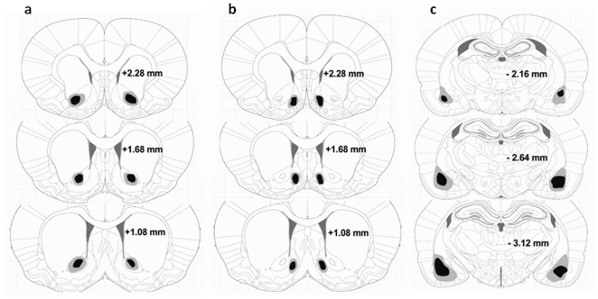
Figure 1. Location of the excitotoxic lesions of the nucleus accumbens core and shell and the basolateral amygdala.
Shown is the extent of the (a) quinolinic lesions of the AcbC, (b) ibotenic acid lesions of the AcbSh, and (c) quinolinic lesions of the BLA. The black and grey areas indicate the minimum and maximum spread of the excitotoxins as assessed by inspection of the slices. Coronal sections are +2.28 mm anterior through +1.08 mm posterior to Bregma (A and B) and −2.16 mm anterior through −3.12 mm posterior to Bregma (C) according to the atlas of Paxinos and Watson [17].

