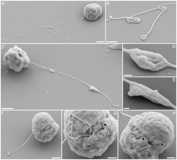
Figure 4. Surface fine structure of male gamete in Pseudostaurosira trainorii and its thread.
SEM. Scales = 2 µm (A, C, F, J), 1 µm (B, G, H) and 0.2 µm (D, E). A. Gamete with branched thread. B. Enlarged view of A showing condensates formed by folded threads. C. Gamete possibly retrieving a thread, judging from the elongated condensates, which are typically seen during thread retrieval. D, E. Enlarged view of C showing elongated condensates unfolding longitudinally (esp. E). F. Gamete winding the thread. G. Enlarged view of F showing a thread irregularly attached to the gamete surface. H. The same gamete as G observed from opposite side. Threads wound around the equator are visible.

