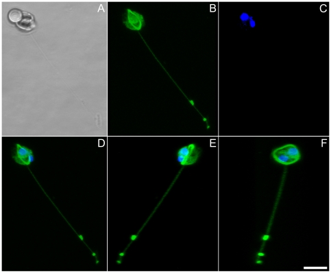
Figure 7. Male gamete in Pseudostaurosira trainorii extruding a thread. LM. Scale = 5 µm.
A. Bright field. B. Tubulin immunolocalization. C. DNA staining with DAPI. D. Merged image of B and C. E, F. Three dimensional reconstruction based on 33 optical stacks taken every 0.21 µm and rotated ca. 130° (E) and 80° (F). Note that a thread stems from the microtubular ring.

