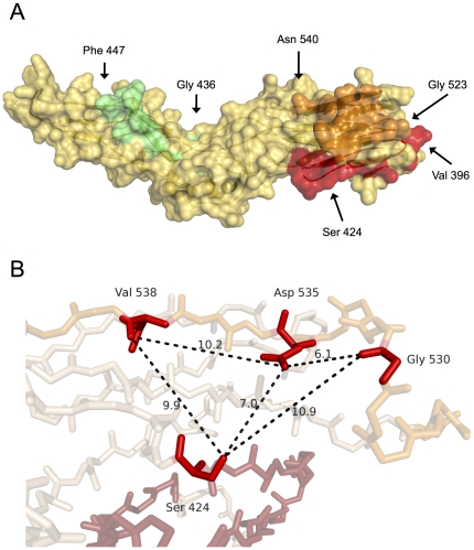
Figure 5. Putative binding site of the AR3B monoclonal antibody on E2.
A. Structural analysis of the proposed AR3B binding site on E2 (orange, red, and green) revealed that it would bury between 1092 and 1794 Å2 at the surface of E2. Regions 396–424 (red) and 523–540 (orange) are closely associated compared with region 436–447 (green). B. Analysis of the 396–424 and 523–540 regions (magnified) showed that critical residues involved in AR3B binding (Ser 424, Gly 530, Asp 535 and Val 538) were largely surface-exposed and lied in close proximity to one another.

