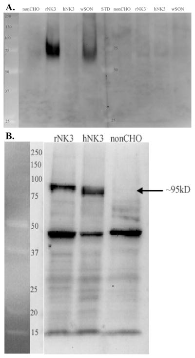
Fig 2.

Western blot of SON and CHO cell homogenates reacted with the N-terminal AB (K2) or the 2nd extracellular loop AB (711). Size standards are shown and labeled (kD) on the left side of each blot. A. The N-terminal AB recognized broad protein bands ranging in size from ~65–75kD in SON and approximately 67–85kD in rat NK3-R transected CHO cells (rNK3). No bands were present in the non-transfected (non-CHO) and human NK3-R transfected CHO cells and both bands were absent when the AB was preblocked with the peptide used to generate the K2 AB (right side of figure). B. The 2nd extracellular loop AB labeled a prominent and distinct protein band ~90–95 kD in both rat and human transfected CHO cells (rNK3 and hNK3) but not in the non-transfected CHO cells. The AB also recognized a protein of ~47kD that is also prominent in the non-transfected CHO cells and therefore, not NK3-R.
