Fig. 3.
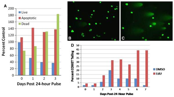
Single-pulse EdU induces delayed cell death and DNA strand breaks. MG63 (human osteosarcoma) cells were treated with a single 24 h pulse of 1 μM EdU or DMSO (vehicle control). a At 0, 1, 2, and 3 days post-pulse, MG63 cells were fixed and immunolabeled with antibody against Annexin V in order to distinguish live, dead, and apoptotic cells. EdU administration demonstrates a progressive increase in dead (green series) and apoptotic (red series) cells at the expense of live cells (blue series) compared to controls. Data are expressed as percent of DMSO-treated cells. b–d DNA strand breaks were detected with the COMET assay at 0–7 days post-EdU exposure. MG63 cells exhibit a progressive increase in cells with DNA strand breakage (c and red series in d) as compared to DMSO cells (b and blue series in d). Images are cells labeled with SYBR® Green. Data are expressed as percent of total cells exhibiting COMET-tailing. Scale bar, 50 μm

