
Figure 3.
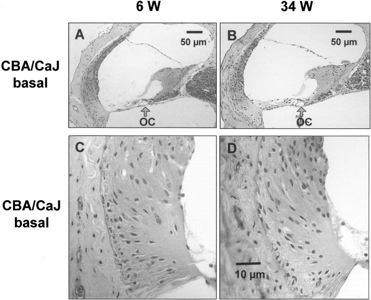
Representative light micrographs of the basal cochlear turn from young and old CBA/CaJ mice. A. The cochlear duct of a 6-week-old mouse (6 W). B. The cochlear duct of a 34-week-old mouse (34 W). Both A and B exhibited almost normal appearance of organ of Corti (OC). C. The basal part of spiral ligament from 6-week-old-mouse (6 W). D. The basal part of spiral ligament from 34-week-old-mouse (34 W). No obvious pathological changes were found in both C and D. The scale bar in D also applies to C.