
Figure 6.
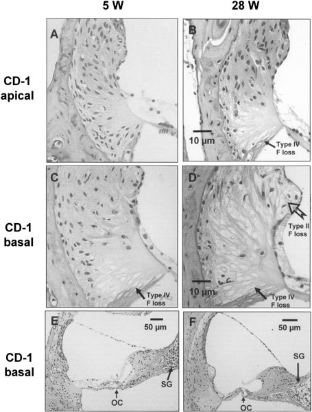
Representative light micrographs from young (5-week-old, 5 W) and old (28-week-old, 28 W) CD-1 cochleas. In the apical turn (A and B at the same scale), there was a loss of type IV fibrocytes (F) at the basal part of the spiral ligament of 28 W (B), compared with that of 5 W (A). In the basal turn (C and D at the same scale), there was a slight loss of type IV fibrocytes at the basal part of spiral ligament of 5 W (C), whereas a more severe and extensive loss of type II and IV fibrocytes in 28 W (D) was observed. In comparison with the normal appearance ofthe organ of Corti (OC) of 5 W basal turn (E), the OC from 28 W basal turn (F) was replaced by simple epithelial cells lacking a distinguishable morphology of the hair cells. The density of the spiral ganglion (SG) in the basal turn of 28 W (F) was clearly lower than that of 5 W (E).