
Figure 7.
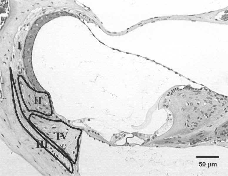
Micrograph of basal cochlear turn of 4-week-old CBA/CaJ mouse demonstrating the locations of type I, II, III, and IV fibrocytes, based on the studies by Spicer and Schulte (1991). The areasof types II, III, and IV fibrocytes are separately represented with labeled outlines and the type I fibrocytes in the rest of the spiral ligament.