
Figure 2.
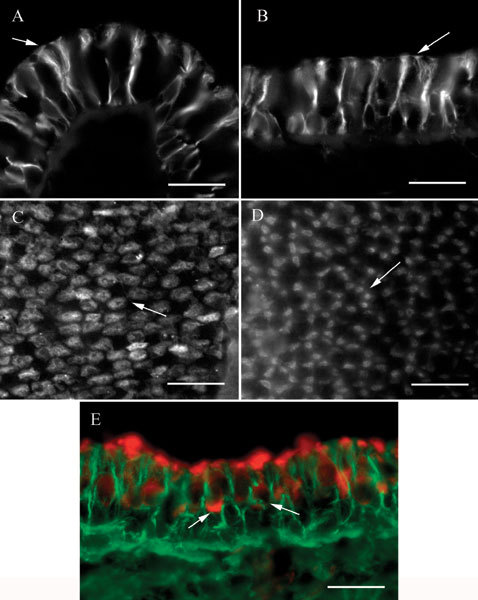
βII tubulin in vestibular epithelia. A Transverse section of a crista ampullaris showing label in supporting cells. Note the large tufts at the apical ends (arrow). B Apical view of a transverse section of a macula. The apical tufts in the supporting cells (arrow) are smaller than those in crista organs. C Apical view of a crista ampullaris whole mount showing the label patches in four or five supporting cells in a cluster surrounding an unlabeled area. A typical cluster is indicated by the arrow. D Whole mount of a utricular macula showing clusters of label patches in the five to seven presumed supporting cells surrounding an unlabeled area. A typical cluster is indicated by the arrow. E. Transverse section of a macula labeled for βII tubulin (green) and myosin VIIa (red). Label for myosin VIIa in two hair cells is indicated by the arrows. The scale bar represents 20 µm for all panels.