
Figure 4.
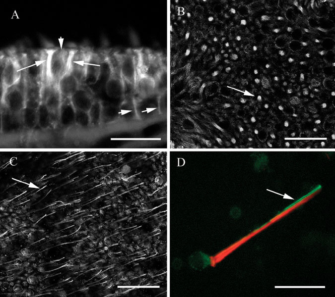
βIV tubulin in vestibular epithelia. A. Transverse section of a utricle showing bright label in the supranuclear regions of hair cells (long arrows) and in supporting cells (short arrows). B Confocal view of a utricle whole mount in a focal plane below the apex showing label in hair cells and supporting cells. The arrow indicates label in a hair cell surrounded by supporting cell label. Confocal image, optical section thickness-0.53 µm. C Apical confocal view of a utricle whole mount showing label in kinocilia (one of which is indicated by the arrow), in hair cells, and in supporting cells. Optical section thickness-0.53 µm. D Isolated vestibular hair cell showing label for actin (phalloidin–red) in stereocilia and the cuticular plate, and label for βIV tubulin (green) in the soma and in the kinocilium. The scale bars represent 8 µm for A and D, 25 µm for B and C.