
Figure 5.
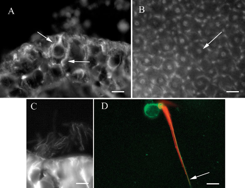
βI tubulin in vestibular epithelia. A Section of a crista showing label in a hair cell and a supporting cell (arrows). B Whole mount of a utricular macula showing patches of label in supporting cells surrounding label in a hair cell (arrow). C. Section of a crista showing labelin kinocilia. D Isolated vestibular hair cell showing label for actin (phalloidin–red) in stereocilia and the cuticular plate and label for βI tubulin (green) in the soma and in the kinocilium (arrow). The scale bar represents 8 µm for A, B, and D and 7 µm for C.