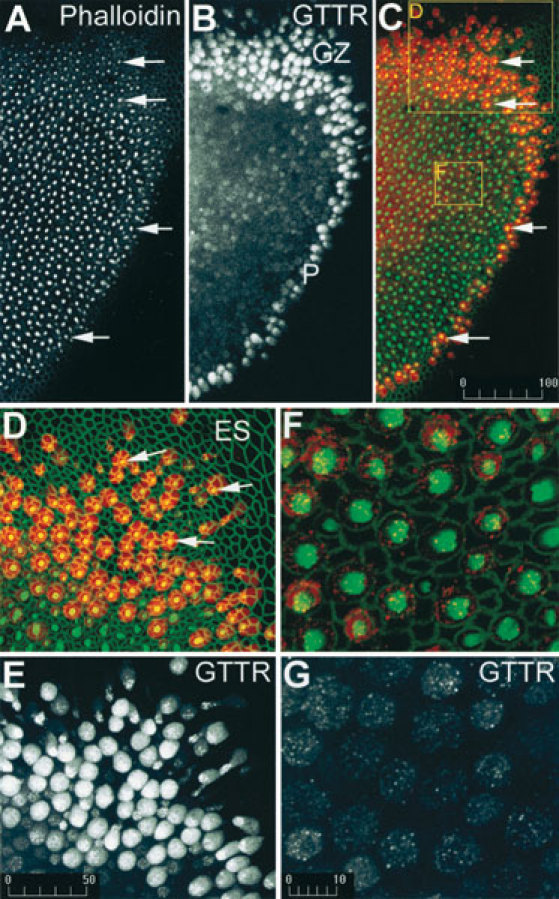
Figure 2.

GTTR is preferentially taken up by hair cells at the periphery of the saccular macula 30 min after addition of 300 µm/ml GT/GTTR. A. At low magnification, FITC-phalloidin labeling reveals a distinct pattern of bright dots (arrows) that represents the sensory hair bundles perpendicular to the surface of the sensory epithelium. B. GTTR fluorescence occurs throughout the sensory epithelium, and most prominently in the growth zone (GZ) and at the periphery (P) of the sensory epithelium. C. A merged image of FITC-phalloidin (green) and GTTR (red), showing the hair bundles superimposed on GTTR-filled cell bodies (arrows). D. At higher magnification, the peripheral red fluorescent cells in the growth zone have intense green fluorescent hair bundles (arrows) at their cell apices. Note negligible GTTR fluorescence in the extrasensory epithelium (ES). E. The red signal only from the image in D reveals negligible labeling of nonhair cells and diffuse fluorescence in hair cells. GTTR fluorescence within peripheral hair cells is both punctate and diffuse. F. In the central region of the saccule, mature hair cells have large round apical surfaces, with an intensely fluorescent actiniferous hair bundle, and are surrounded by the green polygonal outlines of supporting cells. G. The red signal only from the image in F reveals punctate GTTR labeling with diffuse GTTR fluorescence within the hair cell soma only. Scale bars in µm.
