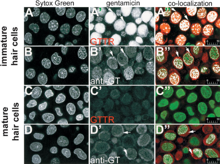
Figure 5.

GTTR and immunolabeled gentamicin are both localized in the nuclei of hair cells. In immature hair cells (A,B), Sytox Green-labeled nuclei appear at the periphery of the sensory epithelium. GTTR (A’) and immunolabeled gentamicin (B’) occur in the same optical plane as Sytox Green-labeled nuclei. Colocalization analysis of single optical planes of nuclei double-labeled with Sytox Green and GTTR (A’’) or immunolabeled gentamicin (B’’) reveal white pixels, indicating nuclei or regions that are colabeled with GTTR or gentamicin antibodies. Only immunolabeled gentamicin (D’’), but not GTTR (C’’), can be readily seen in the nuclei of mature hair cells. GTTR (A’,A’’) and immunolabeled gentamicin (B’,B’’,D’,D’’) is also present in the perinuclear cytoplasm (→). All images are from explants incubated with 300 µg/ml GT/GTTR or unconjugated GT (and subsequently immunolabeled) for 30 min. Scale bars in µm.
