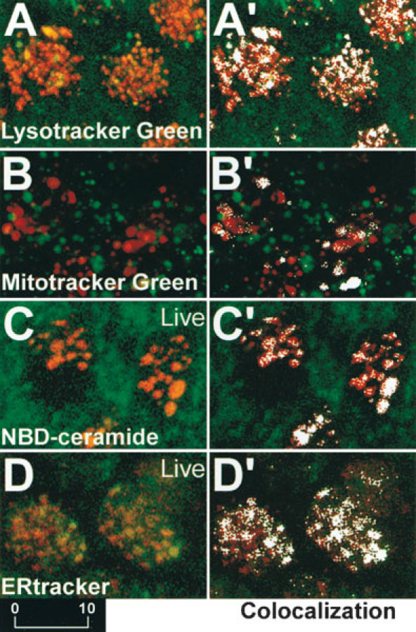
Figure 6.

A–D. Explants preloaded with Lysotracker Green, Mitotracker Green, NBD-ceramide, and ERtracker that fluorescently label lysosomes, mitochondria, Golgi bodies, and ER, respectively, and subsequently incubated with GTTR for 2 h. (A’–D’). Colocalization analysis reveals as white pixels those areas where the red and green fluorescence intensities are above a user-defined threshold, indicating that GTTR is colocalized in the region of fluorescently labeled lysosomes (A′), mitochondria (B′), Golgi bodies (C′) and ER (D′). Scale bar in µm.
