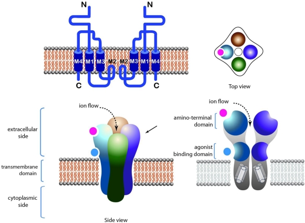
Figure 2.
Structure of tetrameric LGIC. NMDA receptor is taken as an example. Top left: Topology of the receptor. Top right: Top view of the receptor. Bottom left: Side view of the receptor showing the extracellular, intracellular and transmembrane domains. Agonist binding sites are located in juxta-membrane domains in the extracellular side of the receptor. Bottom right: Longitudinal cross section of the receptor, showing the pore domain.

