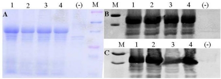
Figure 2.
Detection of expressed fusion proteins. Panel A: Total proteins extracted from inclusion body portion were visualized with Coomassie blue staining. The top and bottom purple markers are 75 and 25 KDa, and the two blue markers inside the purple markers are 50 and 37 KDa. Panel B: detection of refolded fusion proteins by anti-CT antiserum. Panel C: detection of refolded fusion proteins by anti-STa antiserum. The two markers in panel B and C are 50 and 37 KDa. Lane 1: LTR192G-STaE8A; lane 2: LTR192G-STaT16Q; lane 3: LTR192G-STa; lane 4: LTR192G-STaG17S.

