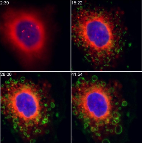
Figure 1.
Expulsion of ricin from intoxicated cells. Cells were labeled with a nuclear dye (blue), and with Bodipy-brefeldin A, which labels ER and Golgi (red). Ricin (green) was added at 3 min post initiation of timing, shown as min:sec in the time series. Within 15 min the ricin has begun to colocalize with the Bodipy-BFA (showing orange). Individual ricin-coated blebs move outward from the cell, most clearly seen between the latter time points.

