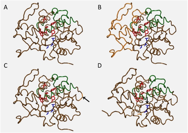
Figure 4.
Crystal structure representations of the recombinant RTA vaccines. RTA active site residue side chains shown in red, VLS residue side chains (L74, D75 and V76) in blue, and dominant immunogenic epitopes in green. A. Wild type RTA; B. RTA1–33/44–198 (USAMRIID) vaccine: portions of the structure genetically excised shown in orange; C. Ricin-MPP (Warwick) vaccine: point of 25-mer insertion shown in orange (with arrow); D. RiVax (Texas) vaccine: Y80A V76M residue side chains shown in orange (Y80A has an arrow). (PDB accession No. 1RTC for panels A, B, and C, 3BJG for panel D.

