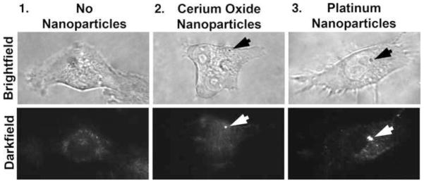
Fig. 2.
Observations of cerium oxide and platinum nanoparticles during interactions with living cells. Nanoparticles were localized using surface plasmon enhanced darkfield microscopy. The nanoparticles are observed as bright dots in the center and right-hand side panels. The resolution of the bright-field images is 240 nm whereas that of the surface plasmon-enhanced darkfield micrographs is 90 nm (Vainrub et al., 2006). (100x objective) (arrows = nanoparticles) (n=5)

