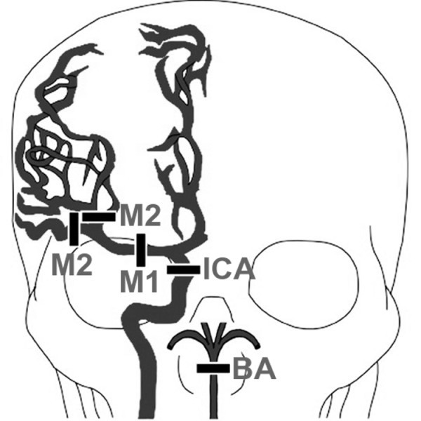
Figure 1.

Large vessel proximal cerebral occlusions. The drawing depicts the major cerebral arteries and the sites of occlusion as specified by the Boston Acute Stroke Imaging Scale classification system (BASIS) [6]. Occlusion sites include the distal internal carotid artery (ICA), proximal segments of middle cerebral artery (M1 and M2), and the basilar artery (BA). Note the exclusion of other proximal arteries including the anterior cerebral, posterior cerebral, and vertebral arteries. The drawing is a modification of the illustration published [6].
