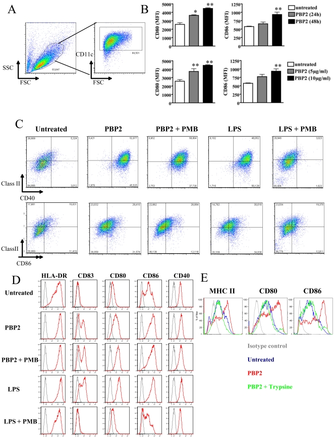
Figure 1. PBP2 induces DC maturation.
A. Gating strategy used in all the experiments using mouse BMDCs. Different staining were then analyzed in the CD11c+ fraction. B: Mouse BMDCs were left untreated or cultured in the presence of 10 µg/ml PBP2 for 24 or 48 hours (upper panel) or with the indicated PBP2 doses for 48 hs (lower panel). CD80 and CD86 expressions were analyzed by FACS. Mean ± SD from three independent experiments is depicted in the graphs. *: p<0.05; **: p<0.01. C: PBP-2-induced phenotypic maturation of mouse BMDCs was analyzed by FACS after a 48 h culture in presence of PBP2 or LPS (from E. coli 0111:B4) alone or in combination with PMB. PMB did not affect PBP2-induced phenotypic maturation whereas it completely abolished LPS- induced expression of CD40, class II molecules and CD86. Numbers in the graphics represent the percentage of double-positive cells. n = 4. D: Human monocyte-derived DCs were treated for 48 h as for C. The expression of the indicated molecules was studied by FACS (red histogram plots). Isotype control staining is depicted in each graphic as grey histogram plots. Cell debris were excluded from the analysis by gating on live cells in the FSC/SSC plot. Representative results from 1 out of 4 donors are depicted. E: PBP2 was left untreated or treated for 1 h at 37°C with trypsin. The enzyme was then blocked with foetal calf serum and preparations were used to treat DCs for 48 hs. One experiment representative of three is shown.

