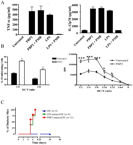
Figure 2. PBP2 increases the immunogenic capacities of DCs.
A: IL-12p70 and TNF-α levels were quantified in the culture supernatant of DCs cultured in the presence of the indicated compounds for 48 hs. A mean ± SD of four experiments is shown. B: Left panel: DDAO-SE-labelled T cells from BALB/c mice were cultured at different ratios for 72 hs with C57/BL6 BMDC previously treated with the indicated compounds. The mean ± SD of divided cells as assessed by DDAO-SE dilution is depicted for two DC/T ratios. n = 2. Right panel: Allogeneic untreated and PBP2-treated human monocyte-derived DCs were γ-irradiated and co-cultured for five days with T cells at different DC/T ratios. Proliferation of responder T cells was studied through analysis of 3H-thymidine uptake. DCs were washed upon PBP2 treatment minimizing the possibility of a direct effect of PBP2 on T cells. *: p<0.05; **: p<0.01; *: p<0.001. C: Diabetes incidence after transfer of untreated (iDC), LPS or PBP2-treated HA-loaded DCs.

