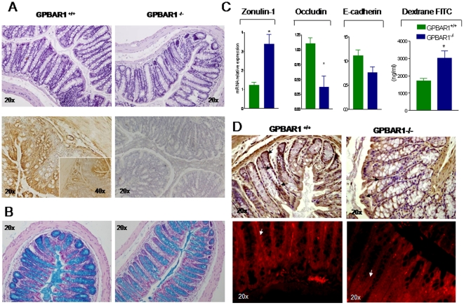
Figure 1. GP-BAR1 gene deletion alters colon structure and function.
Panel A. Upper panels: H&E staining of colonic sections obtained from 12-month old GP-BAR1+/+ and GP-BAR1−/− mice. A significant reduction in colon cellularity and crypt distortion is observed in the colon of GP-BAR1−/− mice. Lower panels: immunohistochemical analysis of GP-BAR1 in wild type mice showing expression in epithelial cells. No staining is observed when the primary antibody is omitted. Original magnification 20× and 40× (insect). Panel B. Alcian blue staining of colon sections from 12-month old mice showing a severe reduction of mucous cells and their impaired maturation in GP-BAR1−/− mice. Note the marked reduction of alcian blue positive cells in the crypts. Original magnification 20×. Panel C. RT-PCR analysis of mRNA expression of junctional proteins (zonulin-1, occludin and E-cadherin) in wild type and GP-BAR1−/− mice (n = 6; P<0.05). 12-months GP-BAR1−/− mice also show a significant increase in intestinal permeability to dextrane FITC (n = 6–8; P<0.05 versus naive). Panel D. Immuno-histochemical detection and immuno-fluorescence localization of zonulin-1. The staining of the protein is increased in GP-BAR1−/− animals but its cells localization appears to be discontinuous (arrows). Original magnification 20×.

