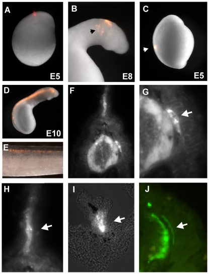
Figure 1. DiI labeling of lamprey neural crest cells reveals absence of sympathetic ganglia during embryonic development.
(A) Focal injections of DiI in the lamprey neural tube at day 5 results in labeling of migrating cephalic neural crest (arrow in B). However, focal injections into the posterior neural tube (C) fail to label trunk neural crest cells. D) Filling the lumen of the neural tube with DiI after cavitation produces labeled trunk neural crest cells in several neural crest derivatives (E). A section through an injected embryo (F) shows labeling of the dorsal root ganglia (DRG, arrow in G), the mesenchyme of the fin (H) and neurons surrounding the gut (F), but no structure that resembles sympathetic ganglia. Neurofilament staining (J) labels neural crest derivatives such as the DRG but also fails to reveal any sympathetic like structures.

