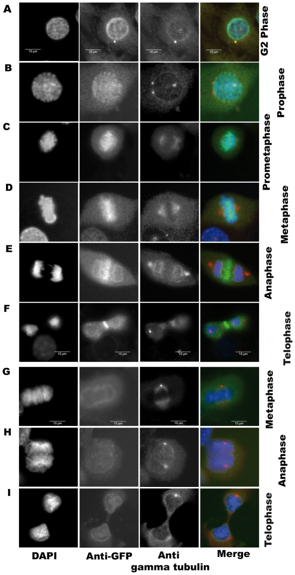
Figure 2. Subcellular localization of GFP-Aurora C along cell cycle.
Immunofluorescence was performed on stable cell clones overexpressing GFP-aurC-WT, GFP-aurC-CA, GFP-aurC-KD and GFP-alone plasmids. Cells were stained with DAPI, anti-GFP and anti-gamma-tubulin antibodies. The immunoflorescent microscopy images show the localization of (A-F) GFP-aurC-WT and (G-I) GFP-alone. Localization of GFP-aurC-WT at (A) centrosome in G2 phase, (B) chromosomes/centromeres in prophase, (C) chromosomes in prometaphase, (D) chromosomes in metaphase, (E) the midzone of spindles in anaphase, (F) midbody in telophase. (G-I) Localization of GFP-alone in (G) metaphase, (H) anaphase, and (I) telophase. The original magnification used was 63×.

