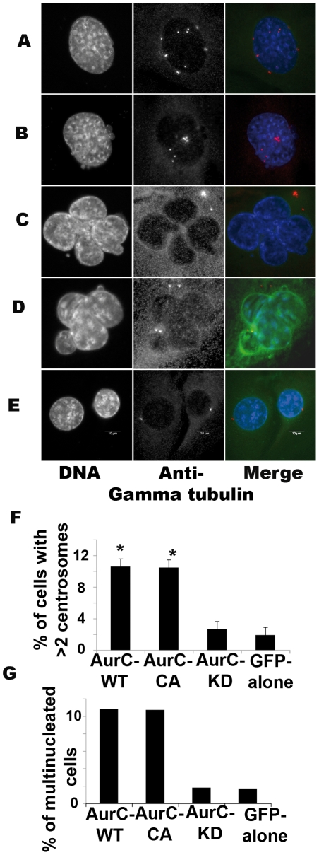
Figure 4. Centrosome number and multinucleation.
NIH-3T3 cells were transfected with (A, C) GFP-aurC-WT, (B, D) GFP-aurC-CA and (E) GFP-aurC-KD vectors, fixed after 96 hours and stained with DAPI and Anti-γ tubulin antibody. (A-B) More than two centrosomes/cell appeared as white dots with anti-γ tubulin staining in (A) GFP-aurC-WT and (B) GFP-aurC-CA transfected cells. (C-D) Multinucleation (more than one nucleus/cell) in (C) GFP-aurC-WT and (D) GFP-aurC-CA in transfected cells. (E) Two centrosomes per cell and only one nucleus/cell in G2 phase of GFP-aurC-KD transfected cells. (F) Histogram shows the percentage of cells with more than 2 centrosomes/cell of 96 hours GFP-aurC-WT, GFP-aurC-CA, GFP-aurC-KD and GFP-alone transfected cells. (G) Histogram shows the percentages of multinucleated cells of 96 hours GFP-aurC-WT, GFP-aurC-CA, GFP-aurC-KD and GFP-alone transfected cells. A minimum of 600 cells was counted for each condition. We performed non-parametric Mann-Whitney test. Results were considered as statistically significant (*) for a p-value under 0.05 when compared to GFP-alone condition.

