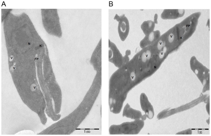
Figure 4. Transmission Electron Microscopic (TEM) images of the A. Wild Type (WT) promastigotes and B. Paromomycin resistant promastigotes.
WT promastigotes were harvested and fixed as mentioned in Materials and methods and subjected to TEM analysis. PRr strain displayed numerous vacuoles compared to the wild type strain. Images are representative of 2 independent experiments. FP: Flagellar Pocket; V: Vacuole; N: Nucleus; K: Kinetoplast.

