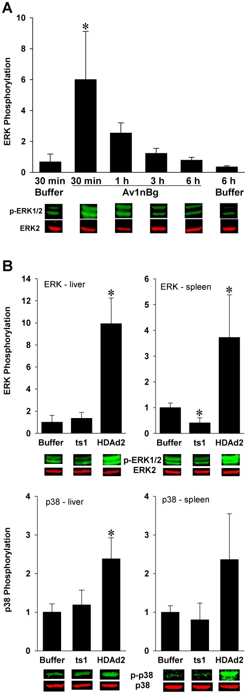
Figure 2. Activation of ERK and p38 by AdV in mouse liver and spleen.
Phospho-ERK (p-ERK1/2) and phospo-p38 (p-p38) were quantitated by Western blot using phospho-specific antibodies. A. Av1nBg, injected i.v. at 5×10¹² vp/kg, induced a rapid phosphorylation of ERK in the mouse liver, with a peak appearing at 30 min. * = p<0.05 vs. 30 min buffer control mice (ANOVA, Holm-Sidak). B. HDAd2 significantly induced phosphorylation of ERK in the liver and spleen and p38 in the liver at 30 minutes, but ts1 did not. Data are normalized to the buffer control group. 5 mice/group. * = p<0.05 vs. buffer control mice (ANOVA, Holm-Sidak).

